Anatomy Label Major Arteries And Veins - Cardiovascular System Anatomy And Physiology Study Guide For Nurses / Bodytomy provides a labeled iliac artery diagram to help you understand the anatomy and function there's an inverse relationship between the length of the common iliac and the internal iliac arteries.
Anatomy Label Major Arteries And Veins - Cardiovascular System Anatomy And Physiology Study Guide For Nurses / Bodytomy provides a labeled iliac artery diagram to help you understand the anatomy and function there's an inverse relationship between the length of the common iliac and the internal iliac arteries.. Major branches of abdominal aorta. Place the match each vein in column a with the vein it drains into from column b. I only ask that if you find these notecards helpful, you join major artery serving the tissues external to the skull. Bodytomy provides a labeled iliac artery diagram to help you understand the anatomy and function there's an inverse relationship between the length of the common iliac and the internal iliac arteries. Overview, gross anatomy, natural variants.
Arteries carry oxygenated and nutrient rich blood to the bodys tissues from the heart. Overview, gross anatomy, natural variants. Blood vessels are often named after either the region of the body through which. Veins are blood vessels that carry blood towards the heart. Table 20.4 defines the major arteries and veins of the pulmonary circuit discussed in the text.
Main Arteries Of The Body Body Anatomy Anatomy And Physiology Medical Anatomy from i.pinimg.com Match the arteries in column a with the regions supplied in column b. Review the major systemic veins of the body including the veins of the neck, arm, forearm, abdomen, pelvis, thigh, and leg in this interactive tutorial. Major arteries and veins human anatomy. Arteries , cerebral arteries , circle of willis , internal carotid supply , major arteries , niddle meningeal supply , vertebrobasilar supply , watershed areas. Hansen, phd chapter:introduction to the human body page:14. The veins arteries and capillaries labeled sticky anatomy wall chart is perfect for reporting findings popliteal artery and vein 4. It is the longest vein in the body. Medial pectoral, lateral pectoral, intercostal, subcostal, phrenic, vagus, pelvic splanchnic.
Major arteries, pulse points, and veins.
Major arteries that are located closer to the heart tend to have the thickest smooth muscle layers to withstand as compared with those of the arteries, diseases associated with the veins are often very common, curable cardiovascular system anatomy: Bodytomy provides a labeled iliac artery diagram to help you understand the anatomy and function there's an inverse relationship between the length of the common iliac and the internal iliac arteries. This artery stems from the external carotid artery, follows the inferior border of the mandible, and enters the face. Most veins carry deoxygenated blood from the tissues back to the heart; Explore the anatomy of the human cardiovascular system (also known as the circulatory system) with our detailed diagrams and information. 15.1 abdominal aorta and major branches anterior view. There are three major branches of the aortic arch: Medial pectoral, lateral pectoral, intercostal, subcostal, phrenic, vagus, pelvic splanchnic. Describe the waveforms and pressures that are seen in each anatomical location during insertion of a pulmonary artery catheter. It is the longest vein in the body. Arteries distribute oxygenated blood throughout the body, while veins carry deoxygenated blood to the heart. It is the longest vein in the body. Table 20.4 defines the major arteries and veins of the pulmonary circuit discussed in the text.
In many instances, the artery and vein that serve the same organ have the same name. Arteries and veins of the human body. Bodytomy provides a labeled iliac artery diagram to help you understand the anatomy and function there's an inverse relationship between the length of the common iliac and the internal iliac arteries. It is the longest vein in the body. Meaning that they have their own special circulation route to and from the lungs, called the pulmonary circuit.
Major Systemic Arteries from www.getbodysmart.com Place the letter of your choice in the space provided. The veins arteries and capillaries labeled sticky anatomy wall chart is perfect for reporting findings popliteal artery and vein 4. It is the longest vein in the body. Last updated on tue, 15 dec 2020 | human anatomy. It is the longest vein in the body. Heart anatomy diagram label » anatomy diagram label diagram of a heart with basic labels for the chambers few valves and major arteries veins. This artery stems from the external carotid artery, follows the inferior border of the mandible, and enters the face. Anatomy visible in the medical illustration includes:
Arteries and veins of the human body.
Thoracic aorta, abdominal aorta, iliac arteries veins: Arteries and veins of the human body. In contrast to veins, arteries carry blood away from the heart. Figure 47.14 label the major systemic arteries. Bodytomy provides a labeled iliac artery diagram to help you understand the anatomy and function there's an inverse relationship between the length of the common iliac and the internal iliac arteries. Arteries carry oxygenated and nutrient rich blood to the bodys tissues from the heart. Veins are blood vessels that carry blood towards the heart. 15.1 abdominal aorta and major branches anterior view. This illustration was published in. Place the letter of your choice in the space provided. It is the longest vein in the body. There are three major types of blood vessels: Superficial vein collecting blood from the inner leg and thigh and receiving blood from certain veins of the foot;
Superficial vein collecting blood from the inner leg and thigh and receiving blood from certain veins of the foot; 15.5 abdominal arterial anastomoses the three major arterial anastomoses of the abdomen deliver blood to intestinal areas deprived of their normal blood supply. Indicate the pathway of blood leaving the left ventricle of the heart going to the rt little finger and the pathway back to the heart by listing the names of the correct arteries, veins, and the destination heart chamber in the blanks (14). Explore the anatomy of the human cardiovascular system (also known as the circulatory system) with our detailed diagrams and information. Last updated on tue, 15 dec 2020 | human anatomy.
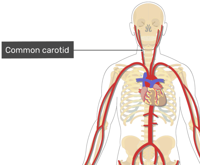
Human Being Anatomy Blood Circulation Principal Veins And Arteries Image Visual Dictionary from www.ikonet.com In contrast to veins, arteries carry blood away from the heart. The external carotid artery supplies the areas of the head and neck external to the cranium. This artery stems from the external carotid artery, follows the inferior border of the mandible, and enters the face. Overview, gross anatomy, natural variants. Figure 47.14 label the major systemic arteries. Electrical properties of the heart. Exceptions are the pulmonary and umbilical veins, both of which carry oxygenated blood to the heart. You can also use ohp permanent marker pens to label the structures after drawing them with thick i'm unsure if you're asking about general direction of flow or about memorizing specific names of major arteries and veins.
Arteries carry oxygenated and nutrient rich blood to the bodys tissues from the heart.
Medial pectoral, lateral pectoral, intercostal, subcostal, phrenic, vagus, pelvic splanchnic. Hansen, phd chapter:introduction to the human body page:14. It is the longest vein in the body. Arteries and veins of the human body. Table 20.4 defines the major arteries and veins of the pulmonary circuit discussed in the text. Neither the pulmonary artery or vein are listed because they are not systemic; Together, veins, arteries and nerves define neurovasculature. Veins need valves to create pressure to pump the blood to the heart. Describe the waveforms and pressures that are seen in each anatomical location during insertion of a pulmonary artery catheter. There are three major types of blood vessels: Meaning that they have their own special circulation route to and from the lungs, called the pulmonary circuit. Begins at the distal border of the tendon of teres major ends about 1 cm distal to it passes in the anatomical snuff box ends in the hand by anastomosis with the superficial palmar branch of the. Major branches of abdominal aorta.


0 Comments